Introduction
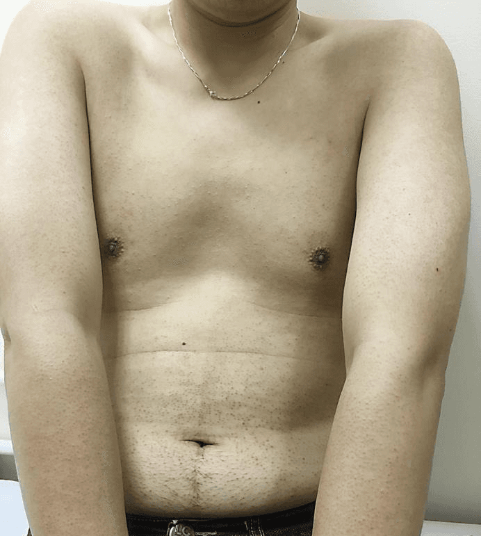
Localized scleroderma (also known as morphea, Latin: morphea) is a rare, chronic inflammatory skin disease characterized by excessive collagen accumulation leading to skin hardening. Although this condition falls within the spectrum of autoimmune diseases, unlike systemic scleroderma, it typically does not affect internal organs. For many patients, the disease is confined to the skin and subcutaneous tissue, but in some cases, it can involve deeper tissues such as fascia or muscles. It is important to know that this disease can be successfully managed, especially with timely treatment and consistent skin care.
Localized scleroderma can impact daily habits, well-being, and self-esteem, which is why the emotional burden of this diagnosis is understandable. Properly selected therapy and regular monitoring by a dermatologist help control disease activity, reduce symptoms, and prevent complications.
What is it?
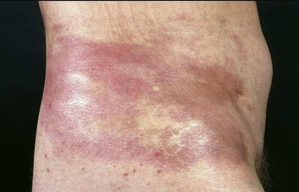
Localized scleroderma is a condition where the immune system stimulates fibroblasts to produce too much collagen. As a result, the skin thickens, hardens, and loses elasticity, and over time, the affected areas may become paler, with an "ivory" hue, sometimes with shine and atrophy. Usually, limited areas of the body are affected, which is why the disease is called "localized."
The most common types of morphea:
- Plaque – one or several round or oval hardened plaques, often in the trunk area.
- Generalized – numerous lesions distributed in different parts of the body.
- Linear – a band-like lesion typical in children and adolescents, often affecting limbs or the face. A linear lesion on the face is sometimes referred to as "en coup de sabre."
- Deep – the lesion involves not only the skin but also the subcutaneous tissue, fascia, and rarely muscles.
- Pansclerotic – extremely rare, affecting large areas and deeper tissues.
The disease course usually begins with an active phase – a visible purple or pink border (inflammatory "halo") around the hardening plaque. Later, the lesion "whitens," becomes hard, shiny, and may eventually thin (atrophy).
Causes and Risk Factors
The exact cause of localized scleroderma is still unknown. It is believed that an imbalance of the immune system, microcirculation disorders, and activation of tissue scarring (fibrosis) processes play a significant role. Some patients have a triggering circumstance before the onset of the disease, but there is usually no single "culprit."
Possible risk factors and triggers:
- Skin injuries, burns, surgical incisions, scars.
- Radiation or previous exposure to radiation in treated areas.
- Certain infections (e.g., after illness), but they do not explain all cases – not every patient has an infectious cause.
- Insect bites or other local irritants.
- Family history of autoimmune diseases or the patient's own medical history.
- Hormonal factors; the disease is somewhat more common in women.
Localized scleroderma is more common in patients with other autoimmune conditions (e.g., autoimmune thyroiditis, alopecia areata, vitiligo, type 1 diabetes). Sometimes it is associated with other inflammatory skin diseases. It is important to emphasize that localized scleroderma is not contagious.
Symptoms
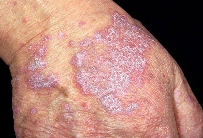
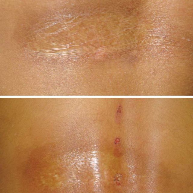
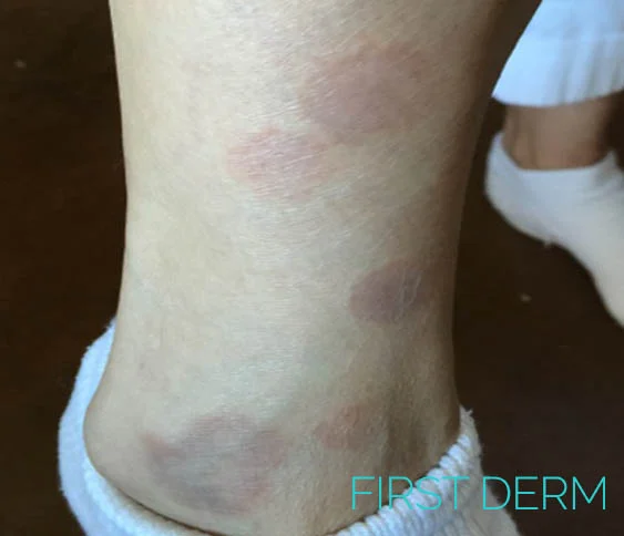
The most common sign is oval or irregularly shaped hardening plaques with a reddish-purple active border. Gradually, they become hard, pale, "waxy," and may easily shine. Around the lesions, pigmentation changes may sometimes be seen: the center is lighter, and the edges are darker. Hair and sweating in the affected area may decrease or disappear completely, and the skin becomes drier and more sensitive to stretching.
Other symptoms and signs:
- A feeling of tightness, sometimes pain or tenderness.
- Restriction of movement if the lesion is over a joint; in the long term, contractures may develop.
- Plaques most often form on the trunk (especially under the breasts, back, or abdomen) but can be on the limbs, face.
- A linear form on the face or scalp can lead to facial asymmetry, thinning hair in the affected area.
- In children, a linear form on a limb can affect growth – the limb becomes narrower or shorter, so early intervention is very important.
Unlike systemic scleroderma, the localized form usually does not cause Raynaud's phenomenon, does not affect internal organs, and does not cause finger hardening. However, extensive or deep disease can impact function and quality of life, especially if the affected areas are over joints or on the face.
When to See a Doctor?
Consult a dermatologist if:
- You notice newly appearing, hardening, color-changing skin spots that do not disappear within a few weeks.
- The plaques are rapidly increasing or new lesions are appearing.
- The lesion is on the face, neck, over joints, or in a child/adolescent – anywhere on the body.
- You experience pain, tightness, movement restriction, or functional impairment.
- You have doubts whether it is another disease (e.g., fungal infection, lichen sclerosus, etc.).
Early consultation allows for a more accurate assessment of disease activity and the selection of an effective treatment plan. Faster initiation of treatment is especially important for the linear form and in children to prevent long-term deformities or growth disturbances.
Diagnosis
The diagnosis is usually made based on the clinical picture and medical history. To confirm the suspicion, a skin biopsy (taking a small piece of skin) is often performed, which, when examined under a microscope, assesses signs of fibrosis and inflammation. Histological examination helps distinguish localized scleroderma from other similar-looking diseases.
Depending on the depth and location of the lesion, the doctor may recommend additional tests:
- Ultrasound of the skin or subcutaneous tissue – to assess depth and monitor dynamics.
- Magnetic resonance imaging – for facial, head, or deep tissue lesions.
- Blood tests – to assess general condition and rule out other autoimmune diseases if symptoms are present.
Our clinic's dermatologists help accurately diagnose, assess disease activity, and develop an individual, scientifically-based treatment and care plan. Consultations are available both in person at the clinic and remotely online – this is especially convenient for monitoring disease dynamics and adjusting treatment.
ARTICLE_GIF
Treatment
Localized scleroderma is not completely curable, but in most cases, it is possible to control its activity, reduce symptoms, and stop progression. The choice of treatment depends on the type, depth, area, activity of the lesion, and the patient's age.
Main principles:
- Topical therapy – special therapeutic creams or ointments for active lesions. They may be prescribed with occlusion (covering) or applied according to a specific scheme to reduce inflammation and soften the skin.
- Targeted injections into lesions – performed by a dermatologist when needed for small, active plaques.
- Phototherapy – light treatment (ultraviolet procedures) under the supervision of a dermatologist. Often used for widespread or multiple lesions to reduce inflammation and improve skin elasticity.
- Systemic therapy – recommended for rapidly progressing, extensive or deep disease, linear form, facial involvement, or functional impairment. The goal is to suppress active fibrosis and protect deeper tissues.
- Physical and occupational therapy – individual stretching, joint mobility exercises, massage techniques, and mobilization help preserve function, especially if lesions are over joints.
- Skin softening agents – continuous moisturizing and strengthening of the barrier reduces tightness, itching, and flaking.
When the disease enters an inactive phase, esthetic dermatology measures (e.g., texture and pigmentation correction) can be considered if safe and appropriate. Disease stability is always ensured first, and then possible cosmetic solutions are discussed.
Treatment effectiveness increases if the patient participates in the process: regularly attends check-ups, follows recommendations, documents changes in lesions (with photos), and informs the doctor of new symptoms. A dermatologist consultation in person or online helps quickly clarify the diagnosis, assess disease activity, and apply a safe, effective treatment plan.
Care and Prevention
Consistent daily care helps protect the skin barrier, maintain elasticity, and reduce discomfort. What you can do yourself:
- Moisturizing – choose rich, fragrance-free products, apply several times a day and always after bathing.
- Gentle cleansing – avoid hot water and harsh cleansers; choose gentle, skin-barrier-friendly ones.
- Sun protection – use broad-spectrum protection daily; affected areas may be more sensitive to UV rays.
- Protection from injuries – wear soft, loose-fitting clothing, avoid prolonged rubbing, pressure, or irritation of affected areas.
- Maintaining mobility – light stretching exercises, especially if the lesion is over a joint; if needed, consult a physiotherapist or occupational therapist.
- Lifestyle – balanced diet, adequate sleep, stress management; psychological support or patient communities can help with emotional well-being.
- Monitoring – periodically photograph lesions in the same light and distance to objectively assess dynamics with your doctor.
The good news is that in most cases, the disease stabilizes over time. However, relapses are possible, so regular dermatologist check-ups help timely adjust care and treatment.
Frequently Asked Questions
- Can localized scleroderma progress to systemic?
For the vast majority of patients – no. These are two separate forms of the disease. However, the doctor evaluates symptoms and, if necessary, orders tests to rule out the systemic form. - Is this disease contagious?
No. Localized scleroderma is not infectious and does not spread from person to person. - How long does the disease last?
The active phase can last months or several years. Later, lesions stabilize, but color or texture changes may remain. Recurrences sometimes occur. - Can children be affected?
Yes. The linear form is more common in children. Early treatment is very important to prevent growth disturbances or movement restrictions. - Is it safe to be in the sun?
Daily photoprotection is recommended. Intense sun can irritate the skin and worsen pigmentation differences, so use protection and wear covering clothing. - Do scars remain after the disease?
Lesions may leave skin thinning or color changes. Aesthetic corrections are discussed only after the disease activity is halted.
Conclusions
Localized scleroderma is a chronic but manageable skin disease. Timely consultation with specialists can reduce inflammation, slow fibrosis, and preserve mobility and well-being. For diagnosis confirmation, a dermato-oncological assessment and, if necessary, a biopsy are often sufficient, and the treatment plan is individually tailored, considering the type, depth, and activity of the lesions. Consistent skin care, maintaining mobility, and regular dermatologist monitoring help achieve the best long-term results. If you notice suspicious skin hardening or color changes, do not delay – the earlier treatment is started, the greater the chance of avoiding complications and restoring skin elasticity.