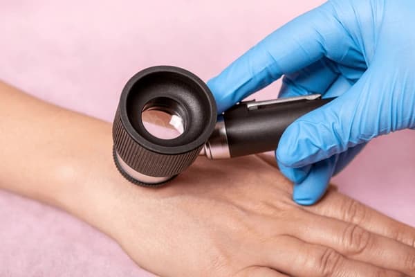
Dermatoscopy – A Method for Examining the Surface of the Skin
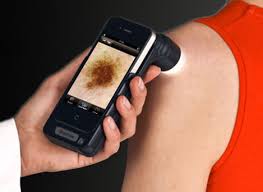
Dermatoscopy is a surface-level skin examination method using a special device called a dermatoscope, which allows for microscopic evaluation of the skin. This method is both informative and easy to apply in daily clinical practice.
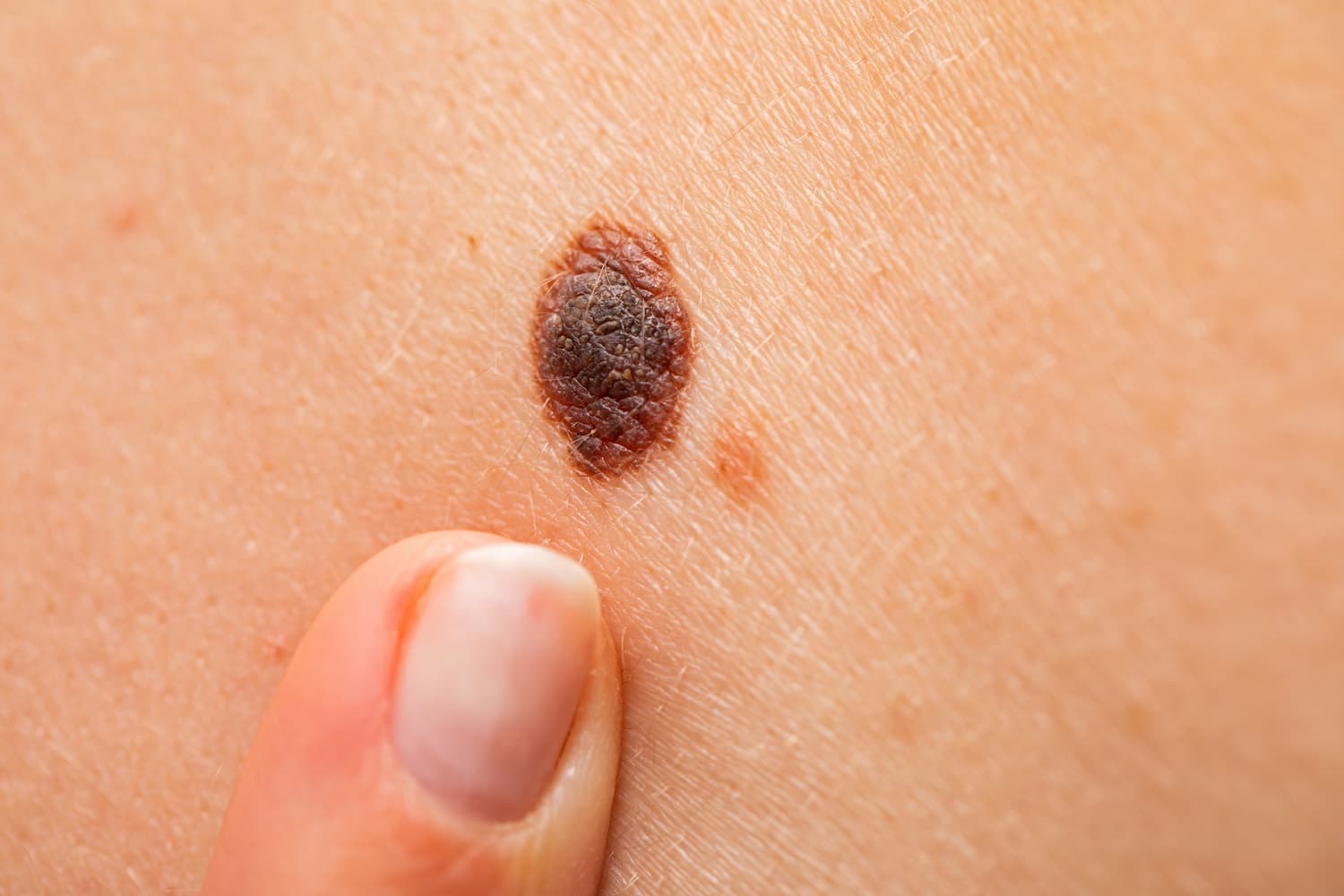
Most commonly, various skin lesions are examined under the dermatoscope. The following aspects of a lesion are evaluated:
-
structure (symmetry, uniformity)
-
color
-
pigmentation
-
the arrangement of blood vessels
-
border characteristics (smoothness, color variation)
-
other changes such as ulceration
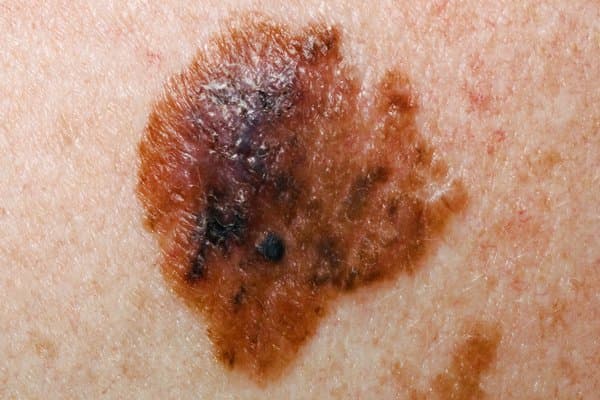
Suspicious features for skin cancer may include:
-
irregular structure
-
uneven color
-
uneven pigmentation, visible changes in pigment tone
-
dense or irregularly shaped blood vessel networks
-
irregular, multi-colored borders
-
visible ulceration or bleeding in the lesion
Using dermatoscopy, it is possible to detect or suspect:
-
melanoma
-
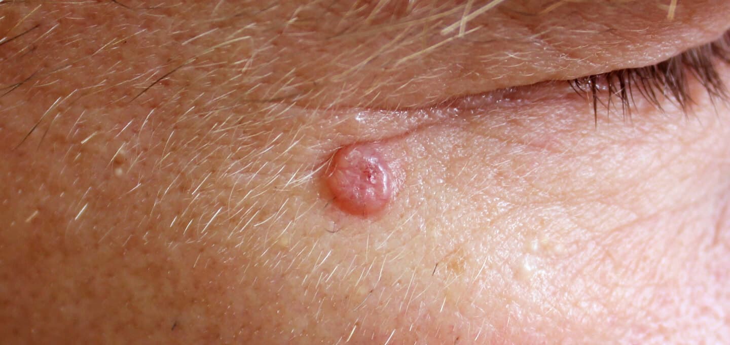
basal cell carcinoma
-
various types of moles
-
seborrheic keratosis
- hemangioma
ARTICLE_GIF
Other uses of dermatoscopy:
-
to determine the type of hair loss (trichoscopy) by examining hair and surrounding scalp
-
to assess non-pigmented lesions
-
to diagnose scabies
-
for differential diagnosis between chronic inflammatory skin diseases, such as:
-
psoriasis
-
eczema
-
lichen planus
-
-
to examine capillaries in the nail bed
-
to locate small embedded foreign bodies, such as splinters