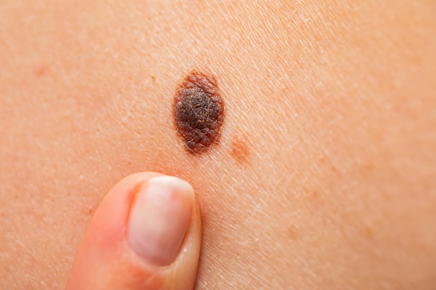
Moles are one of the most common skin features, seen in nearly every person. Most of them are harmless, but it's important to know how to recognize changes that may require medical attention. Benign melanocytic nevi are the most common type of pigmented skin growths, formed from pigment-producing cells called melanocytes. This article explains what they are, what causes them, when to be concerned, and how they can be monitored or treated.
What are melanocytic nevi?
Melanocytic nevi (commonly known as moles) are benign pigmented skin lesions that form when melanocytes cluster in one area of the skin. These moles may be congenital (present at birth) or acquired (developing later, usually during childhood or adolescence).
Causes and risk factors
The number and development of moles are influenced by several factors:
-
Genetics – if parents have many moles, their children are likely to have more as well
-
Skin type – people with fair skin tend to develop more nevi
-
Sun exposure – ultraviolet (UV) radiation stimulates pigment production and mole formation
-
Immune system status – people with suppressed immunity (e.g., due to medication) may develop more moles
Around 1% of people are born with one or more congenital moles.
Types and characteristics
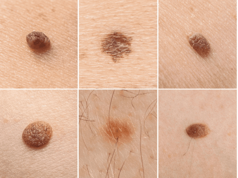
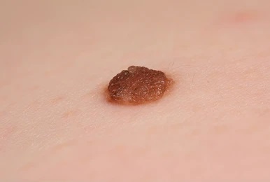
Typical (benign) moles usually appear as:
-
Symmetrical, round or oval in shape
-
Uniform in color – from flesh-colored to dark brown
-
Usually small – up to 6 mm in diameter
-
Flat or slightly raised above the skin
-
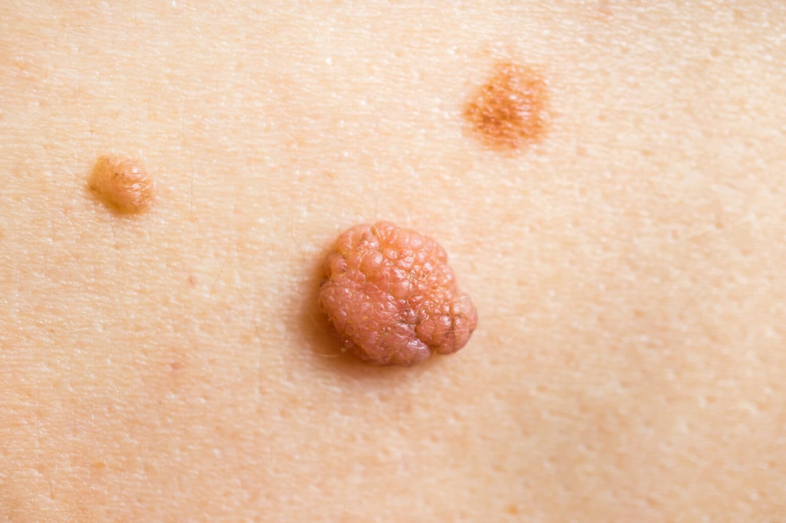
May become lighter or develop a rougher surface with age
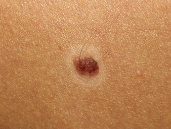
Atypical (dysplastic) nevi may appear as:
-
Asymmetrical with irregular or blurred borders
-
Uneven color – multiple shades of brown, red, or black
-
Larger than 6 mm in diameter
-
Flat or partially raised, often with a rough surface
Having multiple atypical moles increases the risk of developing skin cancer (melanoma).
When should you see a doctor?
You should consult a dermatologist if you notice any of the following:
-
A mole changes in shape, size, or color
-
It begins to itch, bleed, flake, or feel painful
-
The mole is frequently irritated by clothing or shaving
-
There is a family history of melanoma
Early detection is key in preventing complications, especially with suspicious changes.
Diagnosis
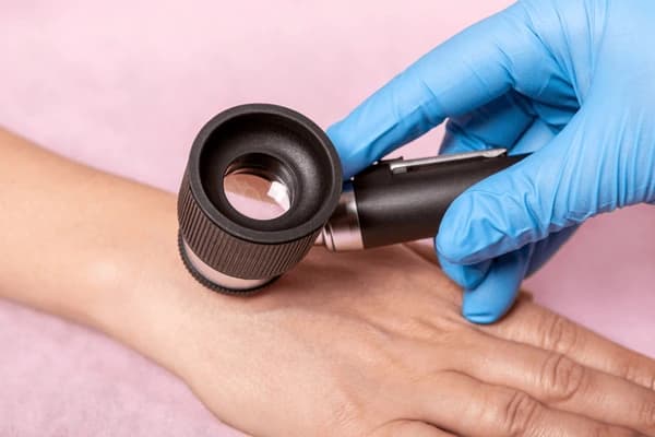
A dermatologist typically assesses a mole visually. For more detailed evaluation, dermatoscopy may be used – a non-invasive method that allows close examination of the mole’s structure and pigmentation.
If cancer is suspected, the following may be recommended:
-
Surgical excision of the mole
-
Histological examination (biopsy) to analyze the tissue under a microscope
Treatment options
Most benign moles do not require removal and can simply be monitored over time. However, treatment may be considered in the following cases:
When mole removal is recommended:
-
Suspicion of malignancy
-
Frequent trauma to the mole
-
Cosmetic concerns (e.g., moles on the face)
Possible removal methods:
-
Surgical excision (sometimes combined with biopsy)
-
Laser removal – suitable for small, cosmetic moles (if cancer is ruled out)
-
Electrocoagulation – destroying the mole with electrical current
Always ensure a mole is benign before removing it for cosmetic reasons.
Prevention and skin care
To reduce the risk of unwanted mole development or progression to melanoma, follow these tips:
-
Avoid direct sun exposure during peak hours (11:00 a.m. – 4:00 p.m.)
-
Use a broad-spectrum sunscreen with a high SPF daily
-
Do not use tanning beds
-
Perform regular skin self-checks – examine your skin once a month using a mirror
-
Visit a dermatologist once a year, especially if you have many or changing moles
Frequently asked questions
Are all moles dangerous?
No – most are harmless. However, monitoring for changes is important.
How often should moles be checked?
Once a year is recommended. If you have many moles or a family history of melanoma, more frequent checks may be needed.
Can moles be removed for cosmetic reasons?
Yes – but a dermatologist should examine the mole first to rule out any malignancy.
How do I know if a mole is suspicious?
Use the “ABCDE” rule:
A – Asymmetry
B – Border irregularity
C – Color variation
D – Diameter over 6 mm
E – Evolving (any change)
If any of these apply, see a specialist.
Conclusion
While most benign melanocytic nevi pose no risk to your health, it's important to keep track of any changes and consult a dermatologist when needed. Regular skin monitoring, sun protection, and early professional evaluation are the best ways to maintain skin health and reduce the risk of skin cancer.