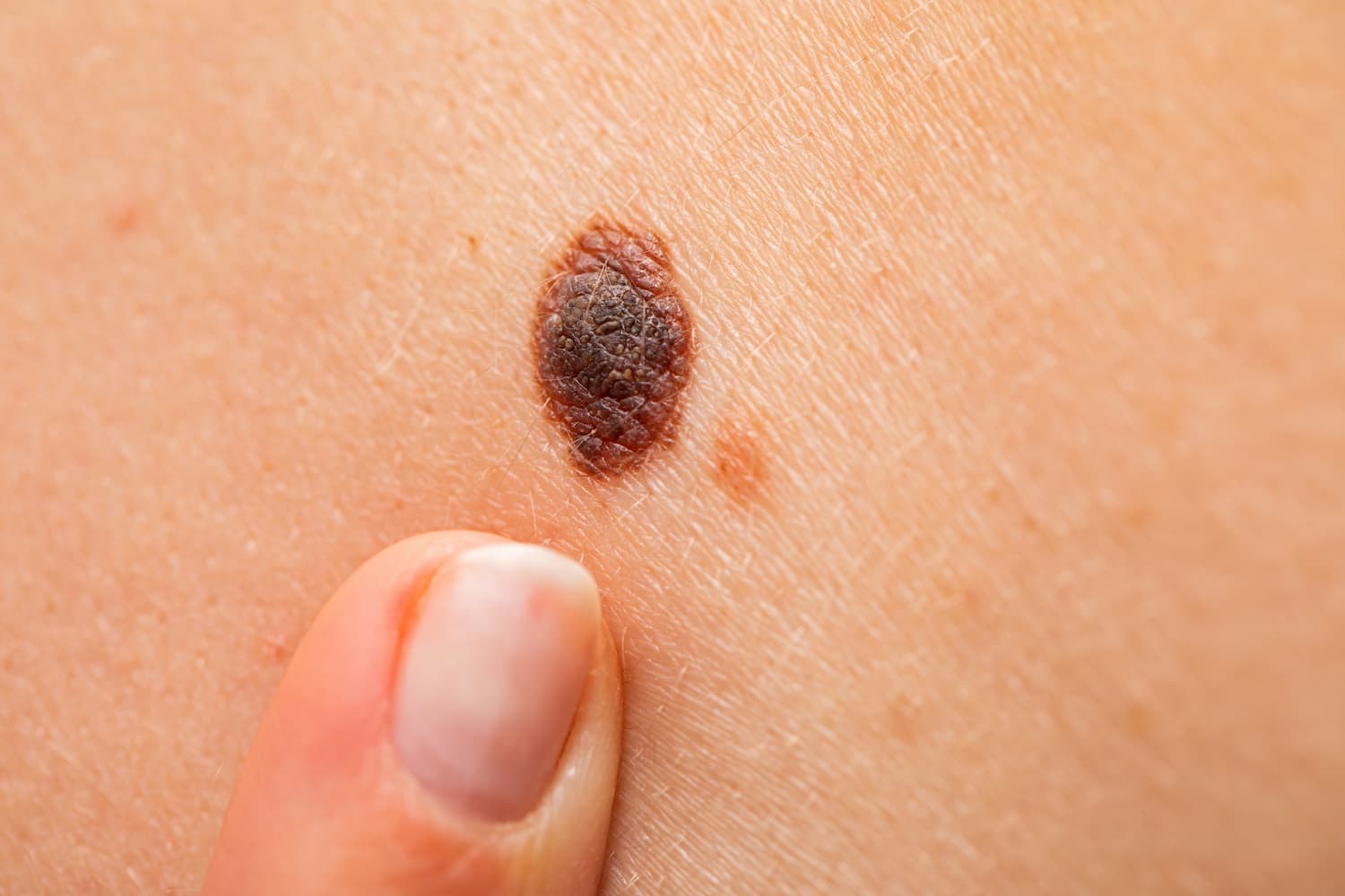
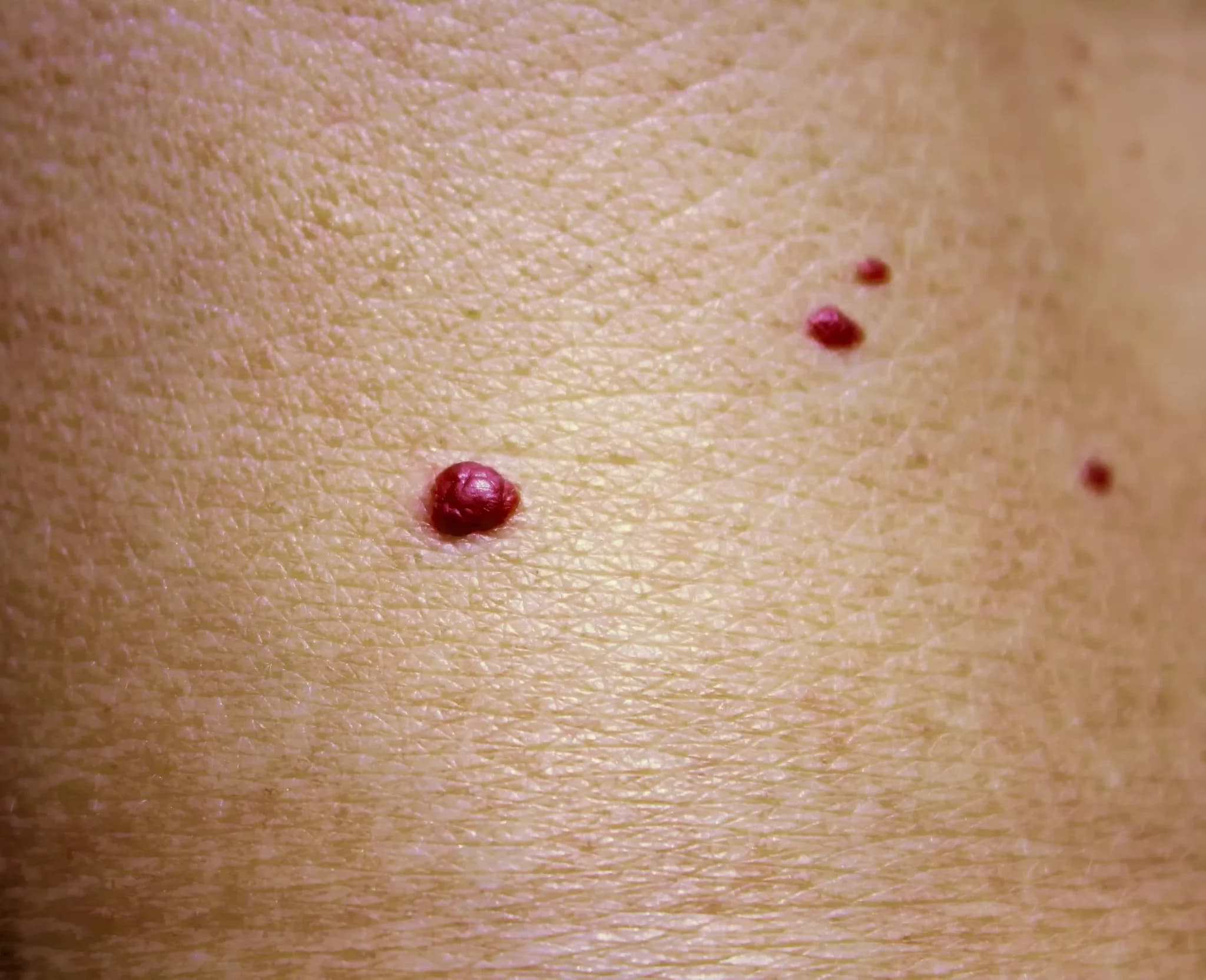
Cherry angioma (also called haemangioma) is a common benign skin growth caused by an overgrowth of small blood vessels. While it usually poses no health risk, its appearance—bright red, dome-shaped, and sometimes multiplying—can raise aesthetic concerns or confusion with more serious conditions. In some cases, cherry angiomas may bleed or appear in large numbers, which is why proper evaluation and, if necessary, treatment are important.
What is a Cherry Angioma?
A cherry angioma is a non-cancerous vascular lesion that forms when clusters of tiny blood vessels (capillaries) grow excessively and become visible through the skin as red dots or bumps. They are composed of proliferated capillary endothelial cells.
Who Gets Them and Why?
-
Most common type of angioma
-
Affects around 7% of adults, and up to 75% of people over 75 years old
-
Usually appears after age 30, but can develop earlier
-
Occurs equally in men and women, regardless of skin tone or ethnicity
-
In rare cases, may be linked to hormonal changes, liver issues, or certain cancers, though not dangerous by itself
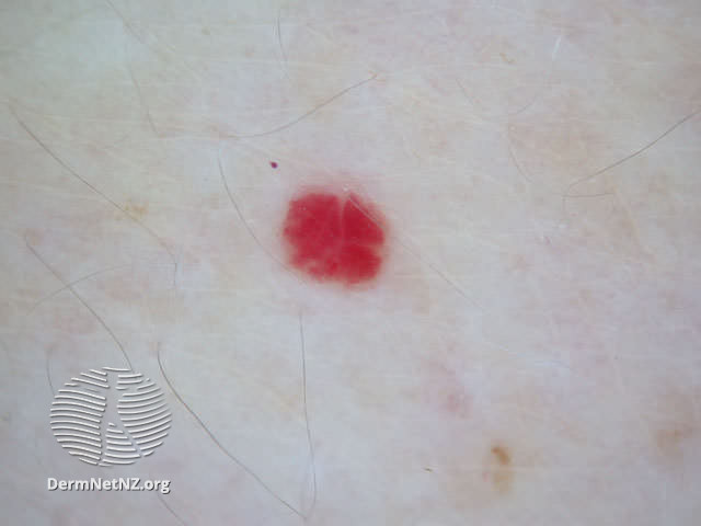

Symptoms and Appearance
-
Begin as small, flat red spots, usually less than 5 mm
-
May grow into dome-shaped papules (raised lesions), up to 1 cm in size
-
Bright red, sometimes purplish or dark
-
Well-defined borders, smooth surface
-
Often appear in clusters, and may merge into larger lesions
-
In some cases, can darken or turn black if a clot (thrombosis) occurs
-
Painless, but may bleed easily when scratched or injured
Most common locations: torso, arms, upper legs, and shoulders. Less often on the face, hands, or feet.
When Should You See a Doctor?
You should consult a dermatologist if:
-
The angioma changes in size, color, or shape
-
It bleeds regularly or becomes irritated
-
It’s in a visible or sensitive area causing discomfort or cosmetic concern
-
You are unsure whether it's benign
-
You experience a sudden increase in the number of lesions
Diagnosis
Cherry angiomas are typically diagnosed through clinical evaluation and visual examination.
Additional tools may include:
-
Dermatoscopy – a close-up look at the blood vessel pattern
-
Biopsy – rarely needed, but used when diagnosis is unclear or to rule out other conditions
Treatment Options
Treatment is usually not medically necessary but may be chosen for cosmetic reasons or if the angioma is prone to trauma.
Common treatment methods include:
-
Electrocautery – the lesion is destroyed using electrical current
-
Laser therapy – precise and effective, especially for small or facial angiomas
-
Cryotherapy – freezing the lesion with liquid nitrogen
-
Surgical removal – used for larger or atypical growths
Prognosis and Monitoring
-
Cherry angiomas are non-cancerous and have no malignant potential
-
They do not go away on their own, but may stop growing or remain unchanged for years
-
New angiomas may develop with age or hormonal changes
-
Once professionally removed, the same angioma does not grow back, though new ones may appear elsewhere
-
Scarring is minimal when removed properly
Frequently Asked Questions (FAQ)
Are cherry angiomas dangerous?
No, they are benign. But sudden changes in appearance or number should be evaluated by a dermatologist.
Can cherry angiomas be removed?
Yes. Safe, effective methods include laser, electrocautery, and cryotherapy.
Do they grow back?
The same angioma won’t return after removal, but others may form in different locations.
How to tell if it’s a cherry angioma or something serious?
Only a dermatologist can confirm the diagnosis. If in doubt, schedule a skin check.
Conclusion
While cherry angiomas are harmless skin growths, they can cause concern due to their appearance or bleeding when irritated. If you're unsure about a red bump on your skin—or simply want it removed for cosmetic reasons—consulting a dermatologist is the best step. Early evaluation helps ensure peace of mind and appropriate care.