Dermatofibroma (also called histiocytoma) is a common benign skin growth that arises from a localized proliferation of macrophages in the dermis. It most often appears on the lower limbs and is seen across all races. Women tend to develop it more frequently than men, typically between the ages of 20 and 50.
In many cases, dermatofibromas develop after minor skin trauma — such as an insect bite, needle prick, shaving cut, or thorn injury. They are also more likely to appear in individuals with weakened immune systems, including those with HIV or autoimmune diseases.
Clinical Features
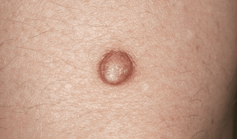
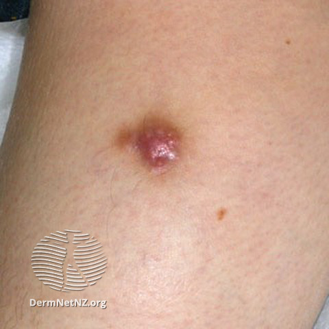
Dermatofibromas typically present as:
-
A firm, slightly raised or flat nodule or papule
-
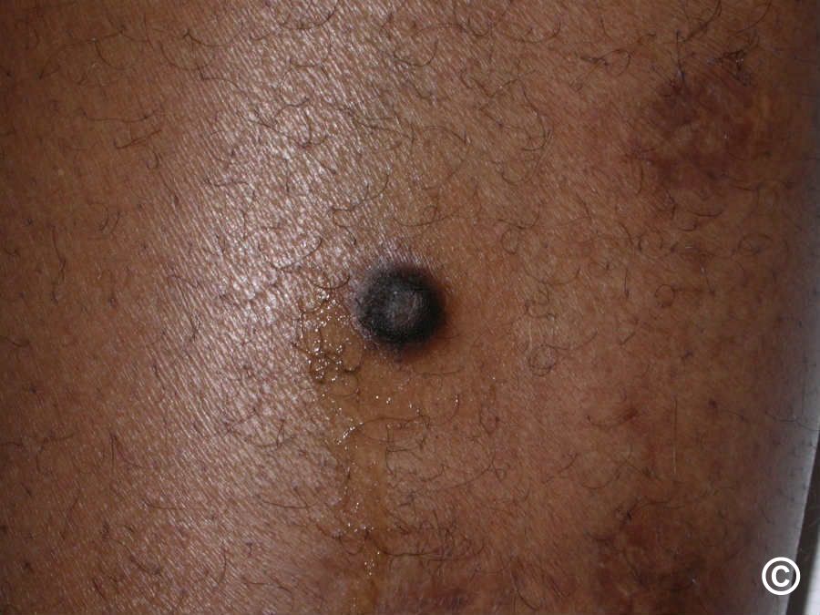
Smooth surface, often pink to light brown in fair skin or dark brown to black in darker skin tones
-
May have a lighter center surrounded by darker pigmentation
-
Size ranges from 0.5 to 1.5 cm, most commonly between 7–10 mm
In most cases, the lesion is asymptomatic, but some individuals may experience:
-
Tightness
-
Pain or tenderness
-
Itching
-
Increased skin sensitivity
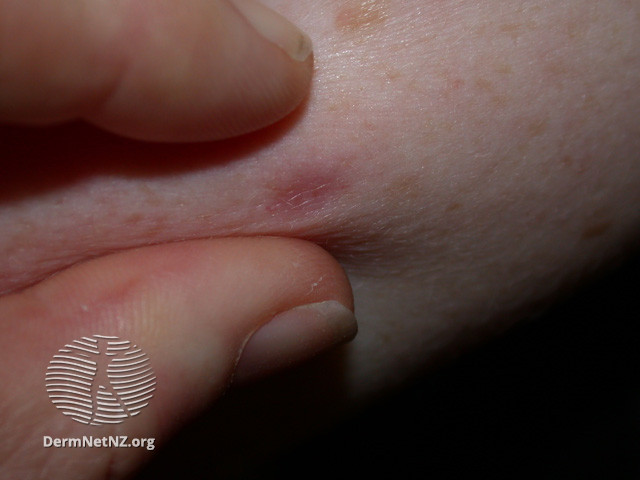
A classic sign of dermatofibroma is the "dimple sign" (also known as the "pinch sign"): when the surrounding skin is gently squeezed, the center of the lesion dimples inward.
Diagnosis
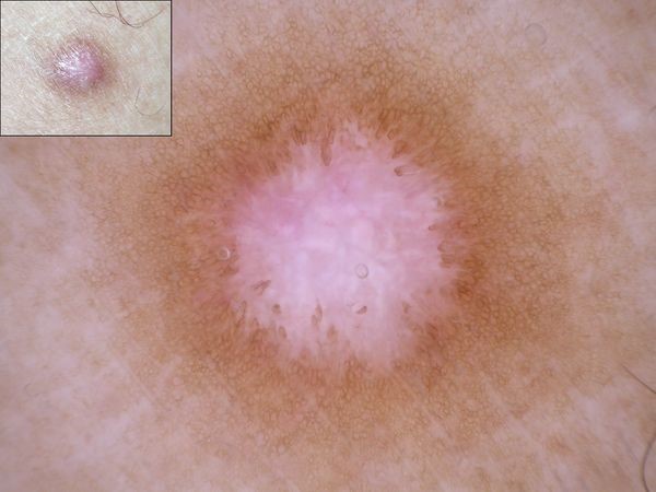
Diagnosis is usually straightforward and made during a clinical examination by a dermatologist. In most cases, dermatoscopy (a magnified skin exam) helps confirm the diagnosis.
In unclear or atypical cases, surgical excision and histological analysis (skin biopsy) may be required to rule out malignant conditions.
Treatment Options
Since dermatofibromas are harmless and non-cancerous, treatment is usually not necessary unless:
-
The lesion causes discomfort
-
There is a need for diagnostic confirmation
-
The patient wishes to remove it for cosmetic reasons
Available removal methods include:
-
Surgical excision (cutting it out)
-
Cryotherapy (freezing treatment)
-
Electrosurgery (using electrical current to remove the lesion)
-
Laser therapy
It's important to note:
-
Recurrence is possible, regardless of the removal method
-
Surgical scars may remain visible and may not meet cosmetic expectations
Important Note
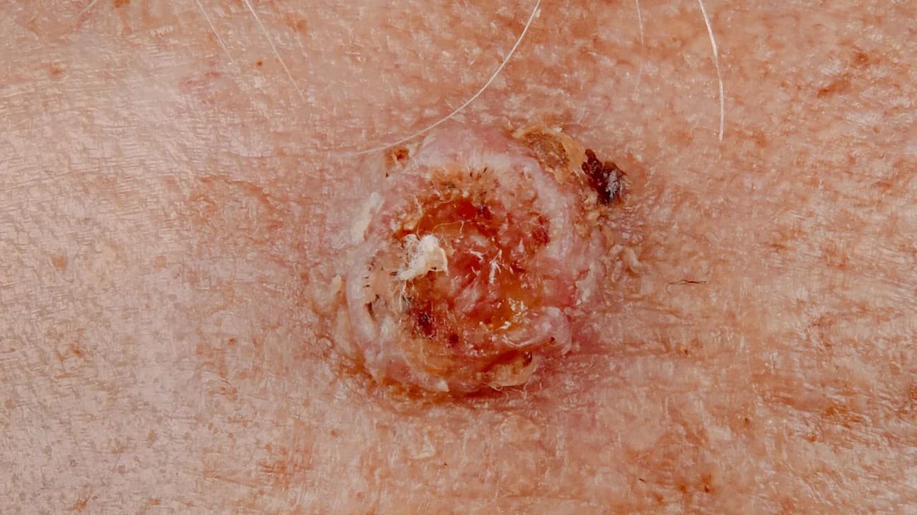
In some cases, dermatofibromas can resemble malignant skin tumors, such as dermatofibrosarcoma protuberans or melanoma. Therefore, any new or changing skin growth should be evaluated by a dermatologist, preferably using dermoscopy. In suspicious cases, a biopsy may be required to confirm the diagnosis.